Introduction
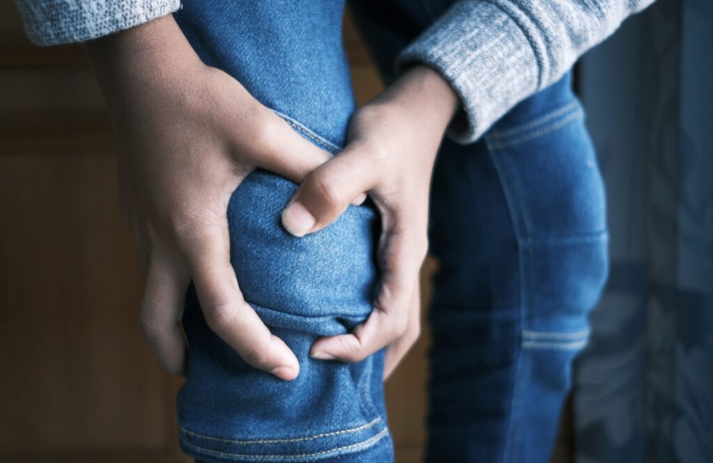
Pain behind the knee cap, also known as patellofemoral pain syndrome (PFPS), is a common musculoskeletal complaint that affects individuals of all ages and activity levels. This condition occurs when the patella (kneecap) does not track properly in the groove of the femur (thigh bone) during knee movements, leading to irritation and inflammation. PFPS can be a frustrating and debilitating condition, affecting daily activities, sports performance, and overall quality of life. In this comprehensive article, we will explore the causes, symptoms, risk factors, diagnosis, and effective treatment options for pain behind the knee cap, empowering readers to take control of their knee health and seek appropriate care.
Anatomy of the Knee and Patellofemoral Joint
To understand the causes of pain behind the knee cap, it is essential to grasp the anatomy of the knee and the patellofemoral joint. The knee is the largest joint in the body, comprising three bones: the femur (thigh bone), the tibia (shin bone), and the patella (kneecap). The patella sits in a groove at the front of the femur and acts as a fulcrum, providing leverage for the quadriceps muscles to extend the leg.
The patellofemoral joint is a unique joint that allows the patella to glide smoothly over the femur during knee flexion and extension. The joint is supported by ligaments, tendons, and muscles, including the quadriceps, hamstrings, and iliotibial band (IT band).
Causes of Pain Behind the Knee Cap
Pain behind the knee cap, or PFPS, can have multiple contributing factors. The most common causes include:
Overuse and Repetitive Movements:
Activities that involve repetitive knee movements, such as running, jumping, squatting, or cycling, can lead to PFPS.
Muscle Imbalance:
Muscular imbalances, particularly weak quadriceps or tight hamstrings, can alter the tracking of the patella and contribute to pain.
Malalignment of the Patella:
Structural abnormalities, such as a misaligned patella or abnormal shape of the femoral groove, can cause the patella to track incorrectly.
Patellar Tendonitis:
Inflammation of the patellar tendon, known as patellar tendonitis or “jumper’s knee,” can lead to pain behind the knee cap.
Knee Osteoarthritis:
Degenerative changes in the knee joint, such as osteoarthritis, can cause patellofemoral pain.
Trauma and Injuries:
Direct trauma or impact to the knee can result in PFPS.
Flat Feet or Overpronation:
Individuals with flat feet or excessive inward rolling of the feet (overpronation) may experience increased stress on the patellofemoral joint.
Tight IT Band:
A tight iliotibial band can pull the patella outward, affecting its tracking.
Symptoms of Pain Behind the Knee Cap

The symptoms of PFPS can vary in intensity and may include:
Pain:
Dull, aching pain behind or around the kneecap, aggravated by activities like climbing stairs, kneeling, or prolonged sitting with knees bent.
Swelling:
Mild swelling around the knee area due to inflammation.
Clicking or Popping Sensations:
Some individuals may experience clicking or popping sensations in the knee during movements.
Stiffness:
Stiffness and discomfort in the knee, particularly after periods of inactivity.
Difficulty Straightening the Knee:
Some patients may have difficulty fully straightening the knee due to pain and tightness.
Weakness:
Weakness in the quadriceps muscles may accompany PFPS, affecting knee stability and function.
Risk Factors for Developing PFPS
Certain factors can increase the risk of developing pain behind the knee cap:
Age and Gender:
Adolescents and young adults, especially females, are more prone to PFPS.
Overuse and High-Impact Activities:
Participating in high-impact sports or activities that involve repetitive knee movements can increase the risk of PFPS.
Muscle Imbalances:
Weak quadriceps or tight hamstrings can affect the alignment of the patella.
Foot and Ankle Issues:
Flat feet, overpronation, or other foot and ankle problems can influence knee alignment.
Previous Injuries:
A history of knee injuries or trauma can predispose individuals to PFPS.
Diagnosis of Pain Behind the Knee Cap

Diagnosing PFPS involves a comprehensive evaluation, including a medical history review and a physical examination. During the examination, a healthcare professional will assess the knee joint, its range of motion, stability, and muscle strength. Additional diagnostic tests may include:
X-rays:
X-rays can help rule out other structural issues in the knee, such as fractures or arthritis.
MRI (Magnetic Resonance Imaging):
MRI scans provide detailed images of soft tissues, helping identify inflammation and tissue abnormalities.
Effective Treatment Options for PFPS
The management of pain behind the knee cap typically involves a combination of conservative treatments, focused on reducing inflammation, improving knee mechanics, and strengthening the surrounding muscles. Some effective treatment options include:
Rest and Activity Modification:
Temporary rest from high-impact activities can help reduce inflammation and allow the knee to heal. Gradual return to activities is recommended, avoiding activities that aggravate symptoms.
Physical Therapy:
Physical therapy plays a central role in the treatment of PFPS. A physical therapist will design an individualized treatment plan, including exercises to improve knee stability, flexibility, and strength. Techniques like patellar taping or bracing may also be used to improve patellar alignment.
Non-Steroidal Anti-Inflammatory Drugs (NSAIDs):
Over-the-counter medications like ibuprofen or naproxen can help reduce pain and inflammation.
Cold and Heat Therapy:
Applying ice packs or using heat pads can provide pain relief and reduce swelling.
Patellar Stabilizing Brace:
A patellar brace is often provided, but has shown to provide very little assistance with return back to sport. If a brace is used chronically, your knee must complete knee stability training by a qualified physical therapist.
Conclusion:
Acute inflammation (inflammation within 1-7 days of initial injury) benefits from P.R.I.C.E.: Protect, Rest, Ice, Compress, and Elevate. However after the acute inflammation phase (essentially after 7 days) exercise and manual therapy is highly indicated for the best return to sport.


